SMS 2022 Image Competition
Imaging in Action
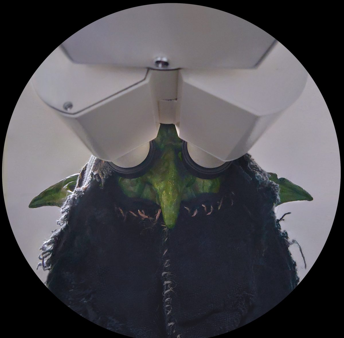
Well done to Dr Kseniia Bondarenko, a Post Doctoral Research Associate at the University of Edinburgh, for their winning entry of ‘Goblinoscope’. Kseniia won the £50 prize
Winner: Goblinscope
Although not all facilities allow this, I bring my inner goblin with me for imaging whenever I can. That particular time happened on October 31, 2022, when some of my Toxoplasma parasites decided to glow red, proudly (and eerily) displaying their flashy mutations! So pretty that they lit up the tumultuous goblin soul, inspiring for an evening of breathtaking images. Optics: Olympus CKX 53 fluorescent inverted microscope. Model organism: Night goblin from the Warhammer universe. Costume: made by myself in between imaging sessions, which we all know by the name of life. Let me know if you’d like to see the full lab goblin story!
Competition Entries
1. Goblinscope
Dr Kseniia Bondarenko, University of Edinburgh
Although not all facilities allow this, I bring my inner goblin with me for imaging whenever I can. That particular time happened on October 31, 2022, when some of my Toxoplasma parasites decided to glow red, proudly (and eerily) displaying their flashy mutations! So pretty that they lit up the tumultuous goblin soul, inspiring for an evening of breathtaking images. Optics: Olympus CKX 53 fluorescent inverted microscope. Model organism: Night goblin from the Warhammer universe. Costume: made by myself in between imaging sessions, which we all know by the name of life. Let me know if you’d like to see the full lab goblin story!
2. Going Through Phases
Dr Charlotte Buckley, University of Strathclyde
Image taken of different modes of a bessel beam created using an SLM in a standard SPIM lightsheet setup. This was taken as part of the initial experiment setup, where we were deciding what mode to use. The homebuilt setup was built at the University of Durham, and we were using using a Bessel beam to very accurately target specific cells in a 3dpf zebrafish to ablate those cells without injuring any surrounding tissue. The lightsheet is incredibly adaptable, allowing changes such as integration of the bessel beam through the imaging objective to adapt the experiment to what you need.

Get in touch with the Scottish Microscopy Society!
Feel free to contact us or join our mailing list using the contact form below.

