Winner of the 2021 'Spotlight on Facilities' Image Competition, sponsored by Zeiss
Competition Entries
1. Abstract art, or aspiring architecture?
Charlotte Clews, The Roslin Institute, Edinburgh
PhD Student
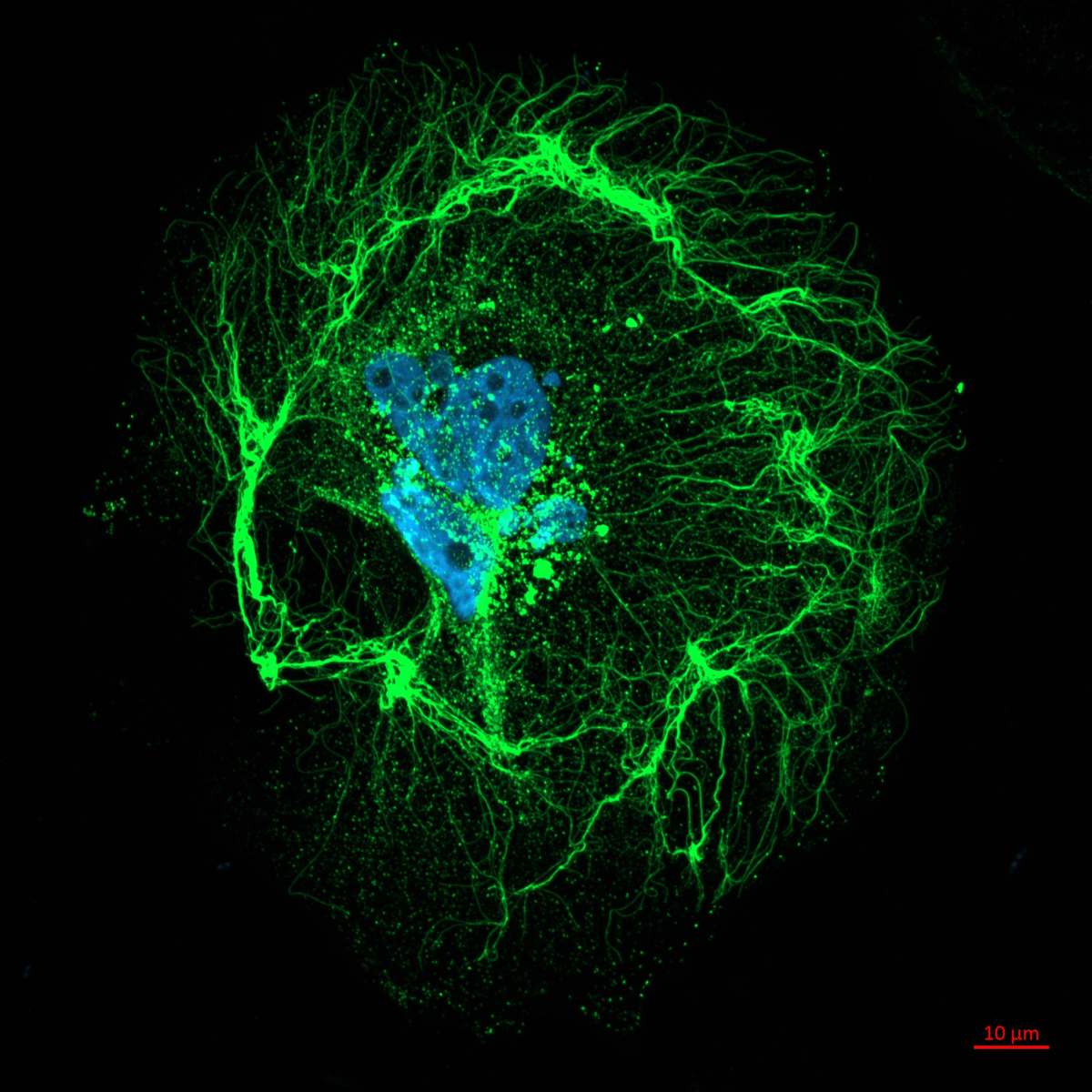
The image shows a particularly picturesque example of collagen deposition, the initial “architectural” stage of hard tissue formation in bone through the production of collagen by osteoblasts before hydroxyapatite mineralisation. The image was acquired while undergoing an MRes investigating the molecular basis of a rare form of Osteogenesis Imperfecta in children which causes defects in collagen deposition and subsequent bone density, of which the cause is unknown.
Represented in the image are two complete stages of collagen production and deposition: firstly the vesicular collagen in the centre of the image, after undergoing ER-Golgi production and trafficking within the osteoblast, is released into the extracellular matrix. We can then observe the successful deposition of this collagen into the 20 – 300 nm diameter fibrils which arch around the cell peripheries. These structures form the collagen scaffold primed for calcium phosphate mineralisation, with this particular image showing the intricacy and artistry of this bio-synthetic process.
2. Neurons at sea
Francesco Gobbo, University of Edinburgh
Post Doctoral Researcher
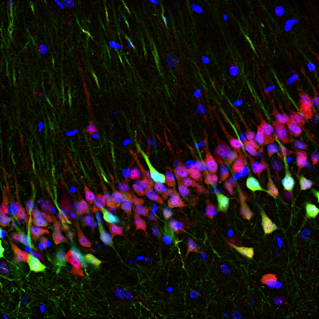
The hippocampus is one of the main brain regions involved in space and memory processing. Here, pyramidal neurons are genetically engineered to express an optical calcium sensor to image their activity in real time with special miniature microscopes.
In this image, the green channel is the endogenous fluorescence of the GCaMP calcium sensor, the red one the anti-NeuN staining visualised with anti-guinea pig Alexa 647 secondary antibody, and in blue is the DAPI staining of cell nuclei.
3. Reconstructing the Vessel
Matthieu Vermeren, University of Edinburgh
Imaging Facility Manager, CRM Imaging Facility
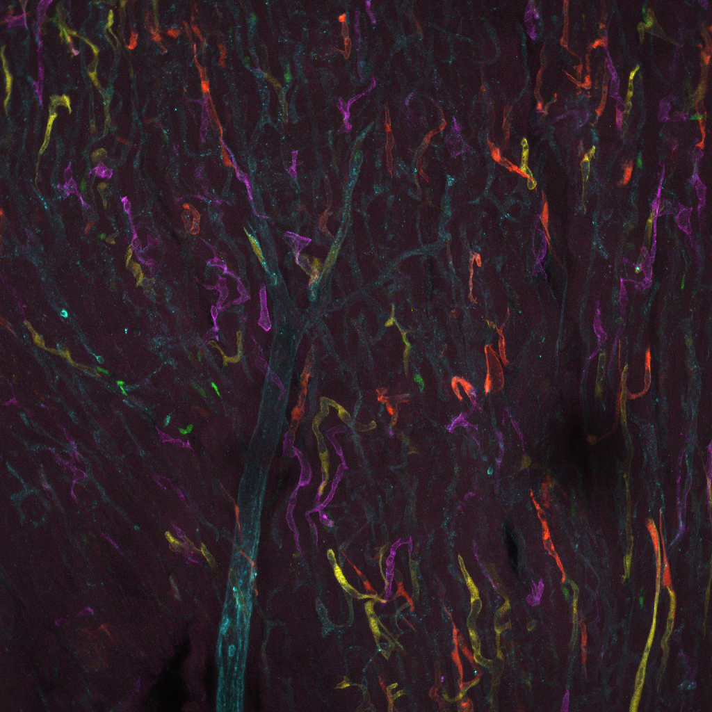
This is a maximum intensity projection of a heart tissue from a confetti mouse imaged on a Leica SP8 with a 25x water lens (CRM, Edinburgh). This tissue has been stained against blood vessels (teal) and nuclei (not shown) and expresses CFP, GFP, YFP, and RFP in different clones. By defining the individual clones by colour and counting their nuclei, the Brittan lab (CVS, Edinburgh) can evaluate how well heart blood vessels grow.
4. Oil Snow Galaxy
Tony Gutierrez, Heriot Watt University
Associate professor of Environmental Microbiology & Biotechnology
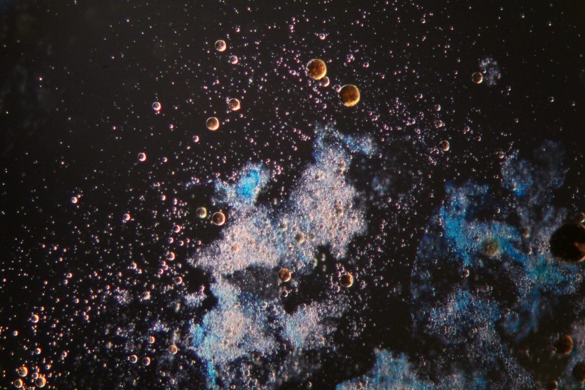
This photo was taken under the light microscope a marine oil snow (MOS) particle stained with Alcian Blue (fluffy blue-white cloud in middle) floating in a sea of oil droplets (brown spheres), of which many were observed embedded within the amorphous matrix of the (MOS) particle. Unprecedented quantities of MOS was formed during the Deepwater Horizon oil spill of 2010 in the Gulf of Mexico, which is thought to have acted as hot spots for the biodegradation of the oil in the water column.
8. Spinning Blood
Emily Munro, Royal (Dick) Schook of Veterinary Sciences, University of Edinburgh
Undergraduate Student
Auto-fluorescing blood cells in mouse tibia stained with phalloidin (a488). Undergoing image analysis in IMARIS at Roslin Institute Bioimaging Facility.
9. Slide Scanning in Action
Lucy Wight, University of Aberdeen
Microscopy & Histology Facility
A brief run through of scanning slides on our Zeiss AxioScan Z1 slide scanner in the Microscopy and Histology Facility at the University of Aberdeen. We use this scanner on a regular basis to scan anything from bone to whale teeth!
10. Rainbow in a Wing
Julia Dong Hwa Oh, The Roslin Institute, University of Edinburgh
PhD Student
Cells in the novel Cytbow chicken line can be induced by Cre-recombinase to express one of three fluorophores: tdTomato, mCerulean and mEYFP. A bead soaked in Cre was inserted into an early wing bud to induce fluorescence in the cells surrounding the bead. The wing was allowed to grow for five more days before dissection and clearing with CUBIC. I travelled to the CVR in Glasgow to use the Zeiss Z.1 Lightsheet microscope (thanks to Colin Loney) and the resulting file was processed with Imaris. My experience with this microscope helped in the acquisition process for Roslin’s own lightsheet (thanks to Anna Raper), which I am very excited to image with further! Our chick wing samples are large, often reaching 1cm in length, and their 3D structure must be intact for anatomical analysis. The lightsheet is ideal for imaging our wings and what is more, it takes less than ten minutes!




Get in touch with the Scottish Microscopy Society!
Feel free to contact us or join our mailing list using the contact form below.


