Winner of the 2019 SMS Image Competition
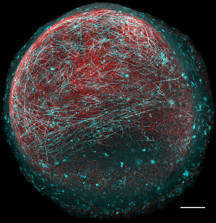
Myelin Moon
Owen James, University of Edinburgh
A human stem cell derived organoid model of myelination with neurons in red and myelin in cyan. Organoids were cultured for 5 months and stained with Abcam 488 (pseudo coloured cyan) against CNP to label oligodendrocytes/myelin and Abcam 568 against NF-H to label neurons. Myelinating organoids can be used study normal and pathogenic myelin development using patient derived iPSCs and to identify new drugs that promote myelin repair. Image was acquired using an ImageXpress spinning disc confocal and is a stitched best projection of over 1000 individual images. Scale = 250µm. Winner’s prize generously donated by ZEISS*.
Runners-up
Anh Hoang Le
CRUK Beatson Institute
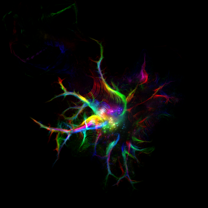
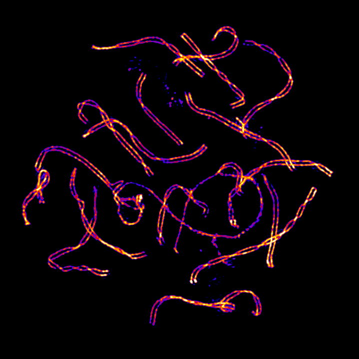
‘The Dancing Cell’. This is a time-lapse image of a melanoma cell. The cell was transfected with LifeAct-GFP and imaged using the Nikon Time-lapse system. Temporal Colour function from ImageJ was applied, so each colour represents a position in time of the cell. This cell is particularly flexible and the curves and waves make it look like it’s dancing.
Ines Boehm
University of Edinburgh
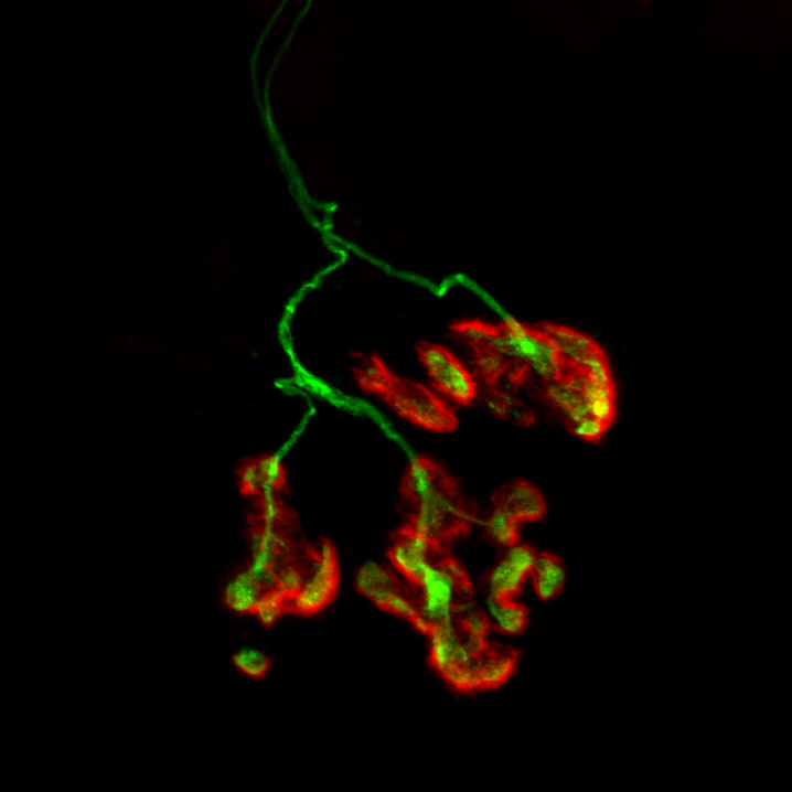
The human neuromuscular junction (NMJ), which is a chemical synapse at the interface between a motor neuron and skeletal muscle fibre and is responsible for signal transmission and subsequent induction of muscle contraction. The postsynaptic terminal in red (TRITC-alpha-Bungarotoxin, 561.8nm laser) depicting acetylcholinreceptors on the muscle fibre and the presynaptic terminal in green depicting synaptic vesicles and the nerve (SV2 and neurofilament, 488nm laser), imaged on a Nikon A1R FLIM, 63x Objective with 3.0 zoom.
Sybille Koehler
University of Edinburgh
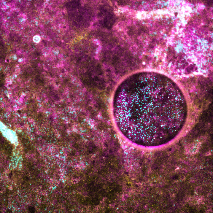
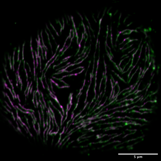
This image depicts the surface of a Drosophila melanogaster nephrocyte.The nephrocyte diaphragm is visualized by antibody staining with Duf (green) and Pyd (magenta) and subsequent imaging with a ZEISS LSM800 and Airyscan.The nephroctye diaphragm is the filtration barrier of the nephrocyte and is the homologue structure to the mammalian kidney filtration barrier.
Dr Katarzyna Styczynska-Soczka
University of Edinburgh
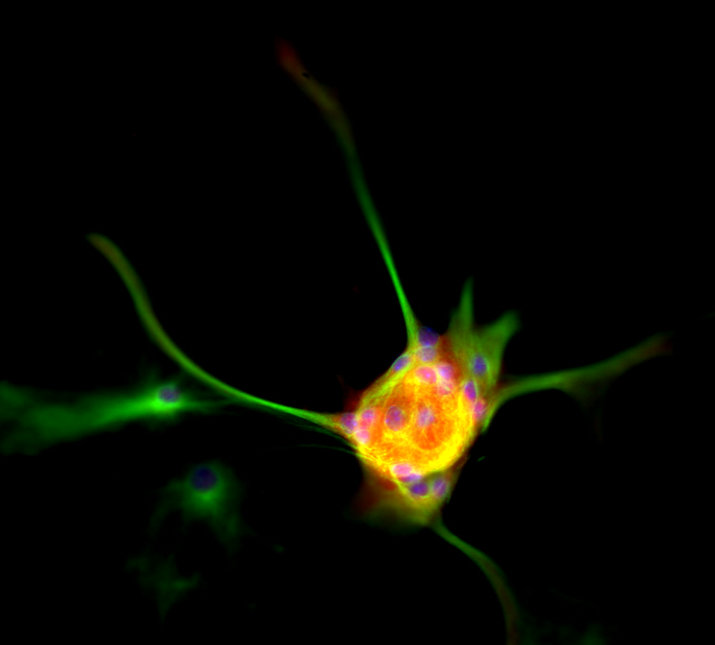
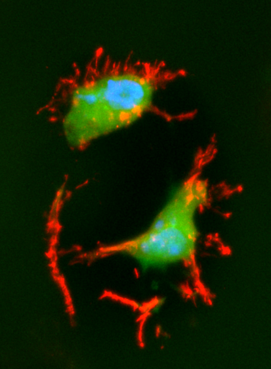
‘Reaching out for help’. Osteoarthritis is a major cause of physical disability and the most prevalent degenerative joint disease in adults. It affects up to 25% of the population over 18 years old and with the ever-growing life expectancy it will be more and more prevalent. This image shows chondrocytes – the only cell type found in the cartilage which covers the end of the bones and permits movement of the joints. Healthy normal chondrocytes are round and have a smooth surface. However, in the early stages of osteoarthritis the morphology of the cells changes. Cells begin to form delicate cytoplasmic processes ranging from single short ones to complex branched networks reaching out far into the surrounding extracellular matrix. Imaged using Zeiss LSM800 confocal microscope, green – CMFDA cell tracker, red – phalloidin (F actin), blue – DAPI.
Amany Hassan
University of Edinburgh
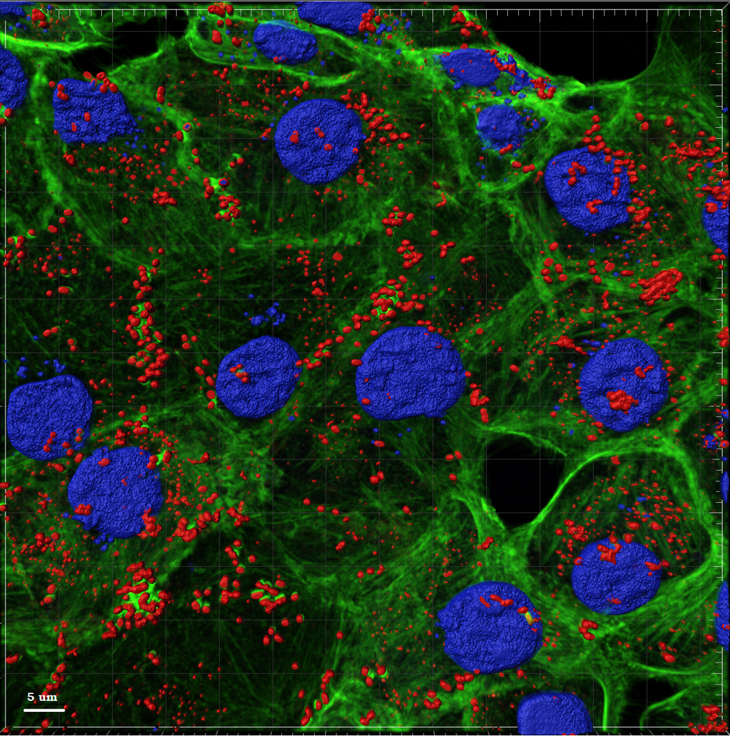
The image demonstrates EHEC O157:H7 colonisation to a bovine epithelial cell line after three hours of infection at MOI of 100. The image shows how the bacteria attach to the epithelial surface with a clear polymerisation of the actin cytoskeleton in green fluorescent color indicating bacterial attachment via T3SS. Image was captured at the Roslin Institute Bioimaging Facility using a Zeiss LSM-880 confocal microscope with plan apochromat x63/1.4NA lens and the following lasers: 405 nm (cell nucleus in blue stained with DAPI), 488 nm (cell cytoskeleton in green stained with phalloidin FITC) and 543 nm (bacteria in red stained with Alexa-flour 568).
*Many thanks to ZEISS for their generous prize (a pair of binoculars worth £250!) for the competition winner.
