Winner of the 2020 Summer Image Competition
Runners-up
Special Mention
Karthik Krishnamurthy
Thomas Jefferson University
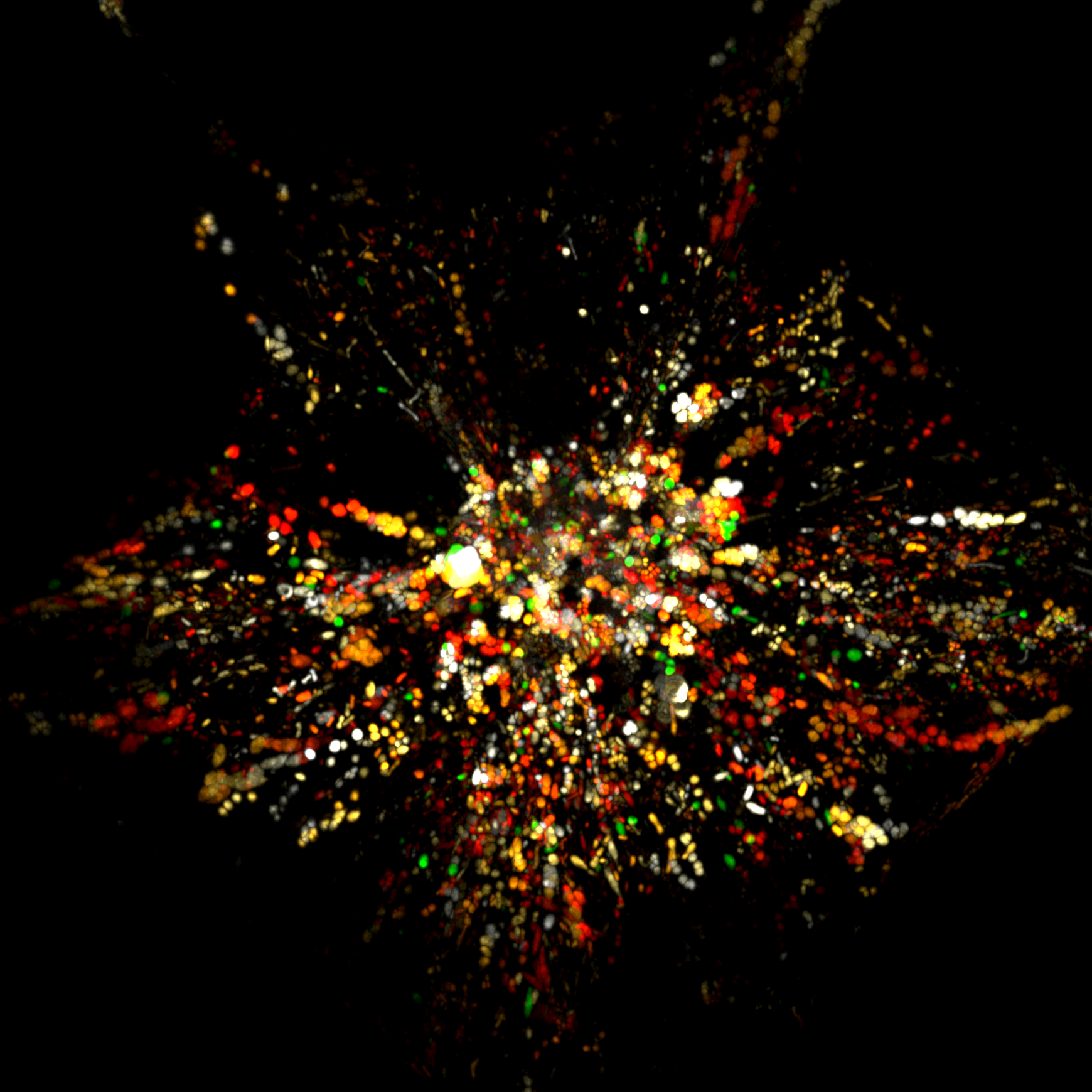
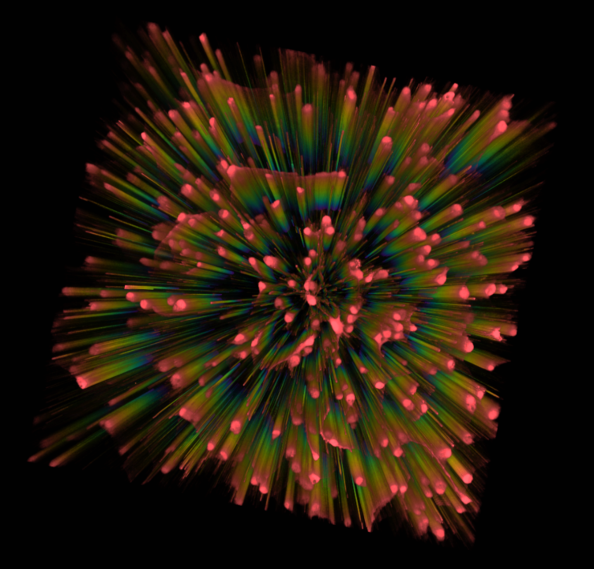
‘Flowering of Neuronal Activity’. Cortical neurons cultured from rat embryos(E17) for 7 days in vitro and labeled with the calcium indicator Fluo-4. Neurons were then stimulated with glutamate and Fluo-4 fluorescence intensity was imaged for 30 minutes at 5 second intervals using a 20X air objective of a nikon Confocal microscope (A1R). Image represents volume view of fluo-4 fluorescence intensity indicative of calcium influx over the 30 minute period blended using depth color coded alpha algorithm as a function of time.
Farah Abdulkhaleq
University of Aberdeen
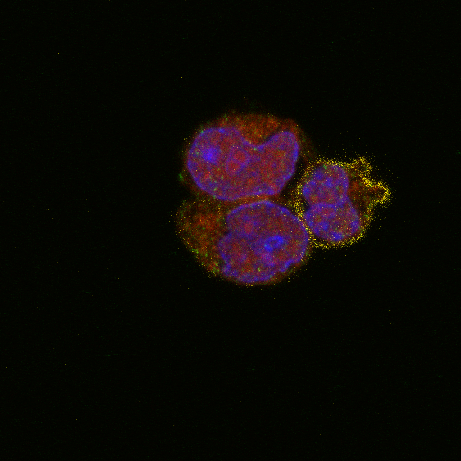
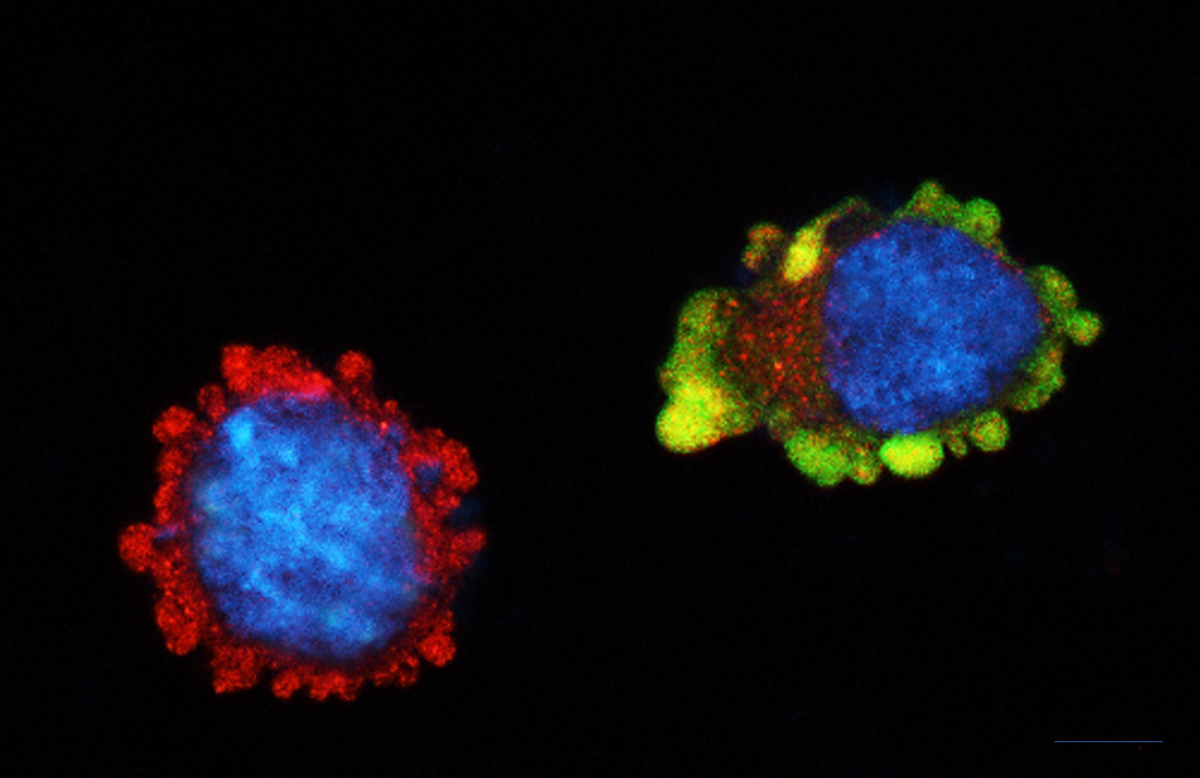
These are 3 stimulated lymphocytes. The one with yellow surface is a CD4+ T cell. Green (goat anti-rabbit IgG (H+L) AF488) and red (streptavidin AF555) channels are CTLA-4 and sCTLA-4, respectively, while the blue channel (DAPI) represents the nucleus. Image was taken using confocal laser scanning microscopy (LSM 880).
The funny thing is that the cells together form the shape of a bee!
Azita Kouchmeshky
University of Aberdeen
NSC-34, a mouse motor neuron-like hybrid cell line, transiently transfected with the (GFP)-SOD1G93A going through autophagy process near a non transfected cell in autophagy phase labelled by LC3B marker. Image acquired using a Nikon field fluorescence microscope)
Simon Brown
University of Edinburgh
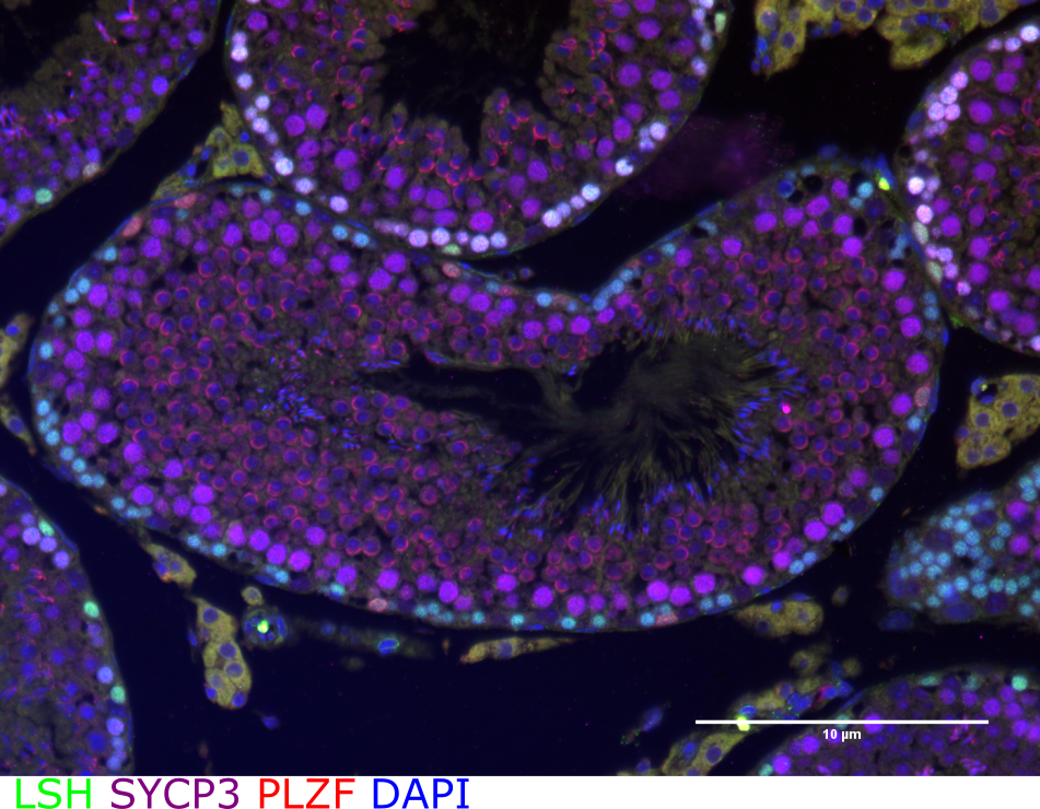
PFA-fixed cross section of adult mouse testes stained with lymphoid specific helicase (LSH), promyelocytic leukemia zinc finger (PLZF), Synaptonemal Complex Protein 3 (SYCP3) and DAPI conjugated to Alexa Fluor secondary antibodies (Fluorophores 488 LSH, 594 PLZF, 647 SYCP3).
Madara Brice
University of Edinburgh
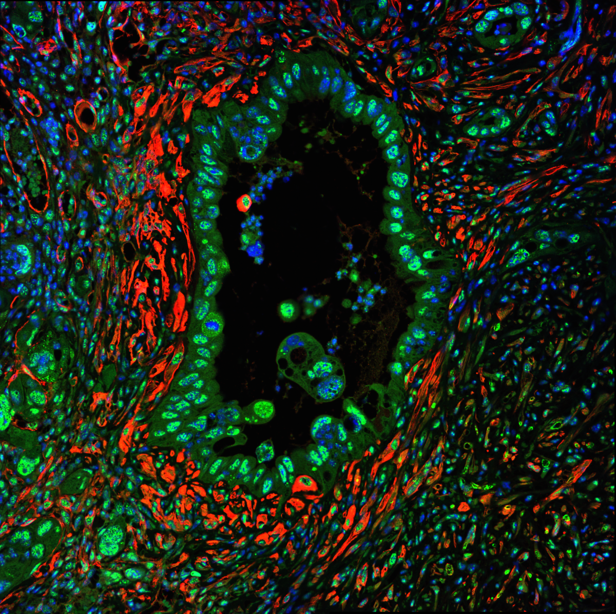
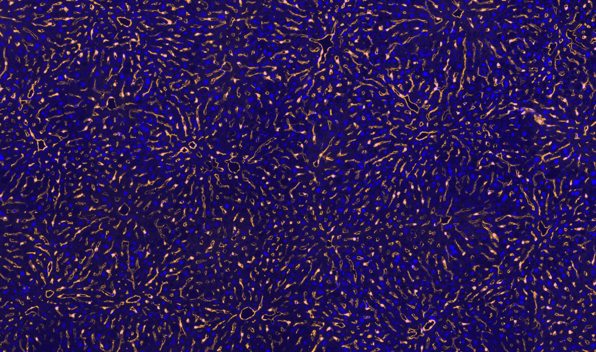
Cross-section of the liver of a c-Kit Cre reporter mouse, although the reporter is actually labelling all endothelial cells (as apposed to sub section with c-kit positivity). Yellow stain – cytoplasm of all endothelial cells, blue – DAPI. Picture taken on an AxioScan Slide Scanner.

