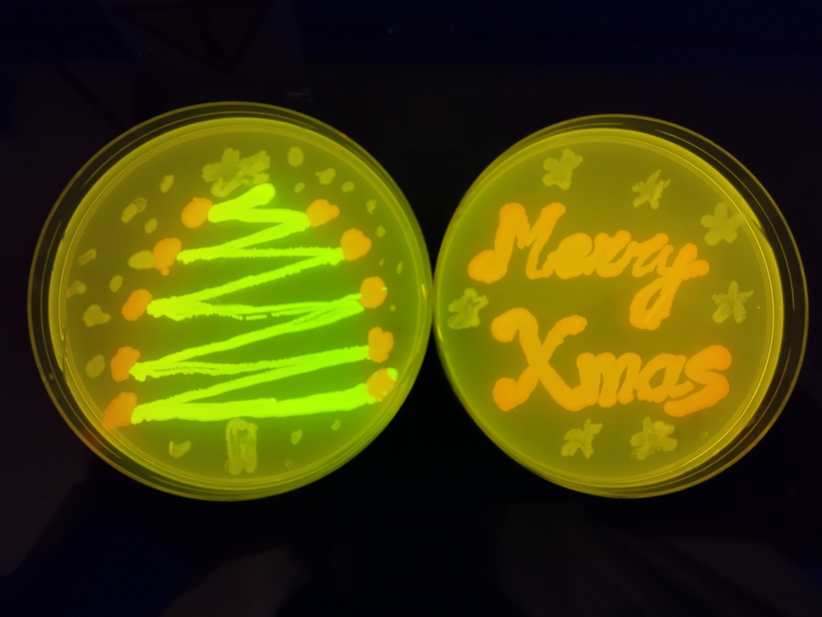
Well done to Dr Jamie Whitelaw, a Lecturer at the University of the West of Scotland and CRUK Beatson Institute, for their winning entry of ‘Cells Actin like a Xmas Tree’. Jamie won the £50 prize
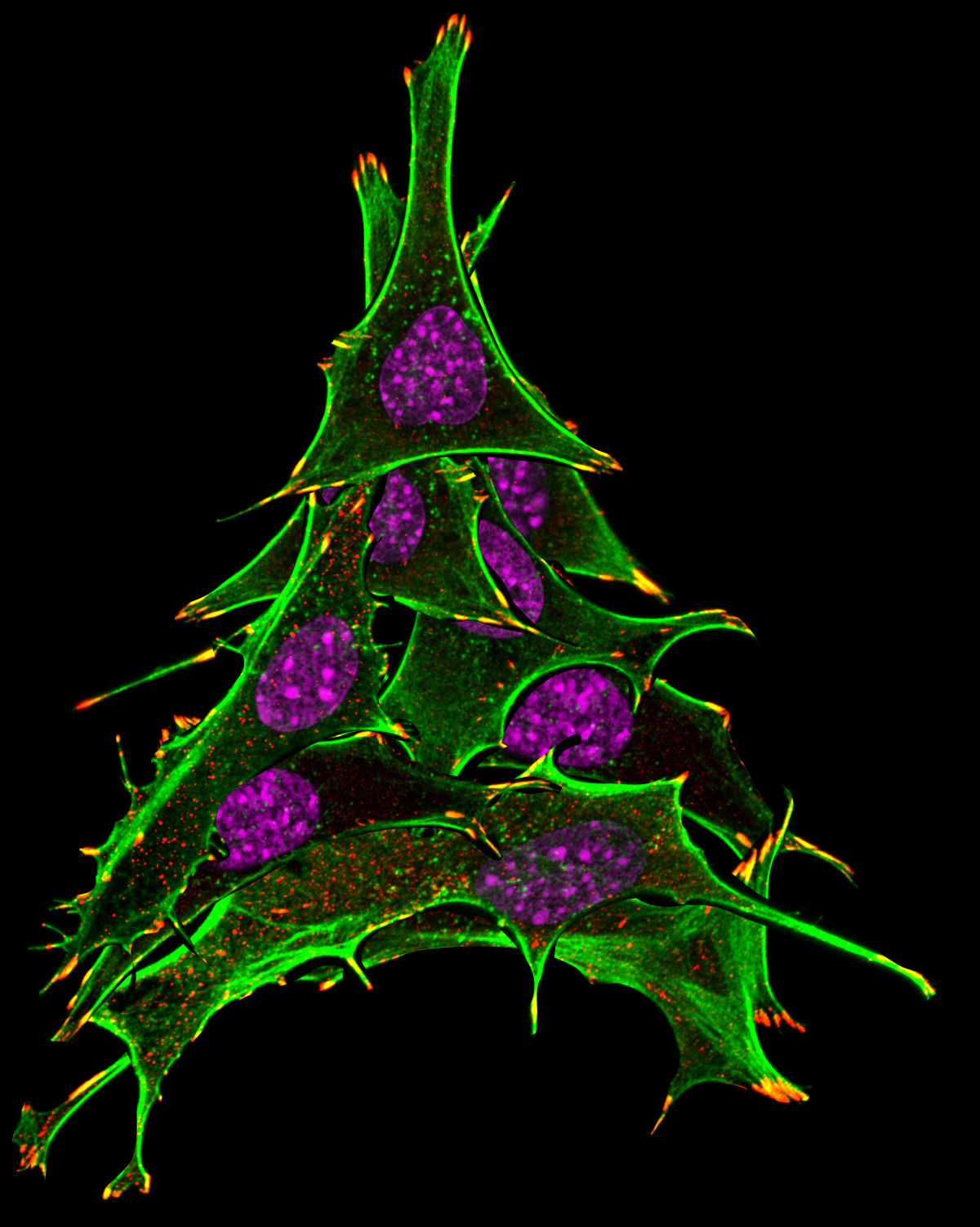
Winner: Cells Actin like a Xmas Tree
Dr Jamie Whitelaw, University of the West of Scotland/CRUK Beatson Institute
NckAP1 KO MEFs stained with Vinculin (Red), Phalloidin (Green) and DAPI (Magenta). These were imaged on the Zeiss LSM 880 at the Beatson BAIR facility using the 63X oil objective lens. The images were processed to form an Xmas tree where the actin staining is the tree, the DAPI is baubles and the Vinculin as the Xmas lights.
Competition Entries
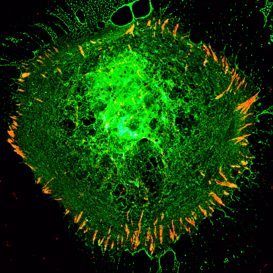
1. The Christmas Star
Charlotte Clews, The Roslin Institute, University of Edinburgh
Osteoblast cell line transfected with fluorescent PHOSPHO1-mNeonGreen fusion construct, and probed for vinculin (indirect immunofluorescence). This shows the activity of PHOSPHO1 within the cell, and vinculin prescence at the periphery of the cell, suggesting the presence of focal adhesion complexes. These points also seem to be points of release for small vesicular objects positive for PHOSPHO1, thought to be matrix vesicles, which are essential for the initiation of calcium phosphate based mineralisation. But I also think this particular cell looks like a star, perhaps a supernova? At least the christmas star wasn’t that dramatic…!
2. Vascular Calcium during Regeneration
Jade Phillips, CRUK Beatson Institute
Genetically encoded calcium signal (GCamp7s- GFP) in the Drosophila tracheal system (TdTomato) which is a direct homologue of the mammalian vasculature during infection induced intestinal regeneration. The video was produced using an ex vivo culture of the entire Drosophila gut capturing only the R4 region of the posterior midgut. A time series was captured over 6-16 hours post infection using a x20 magnification at a Zeiss 780 confocal microscope in the Wolfson Wohl Centre for Cancer Research.
3. Christmas Lights
Dr Chiara Pirillo, CRUK Beatson Institute
Immunofluorescence Ce3D multiplex volume imagine of precise-cut mouse lung section. Tissue was stained for CD4 (blue and green), MHCII(magenta),CD11c(deep orange), B220(red) and CD68(bright orange).The image was taken using a Zeiss LSM880 confocal microscope with lamba mode
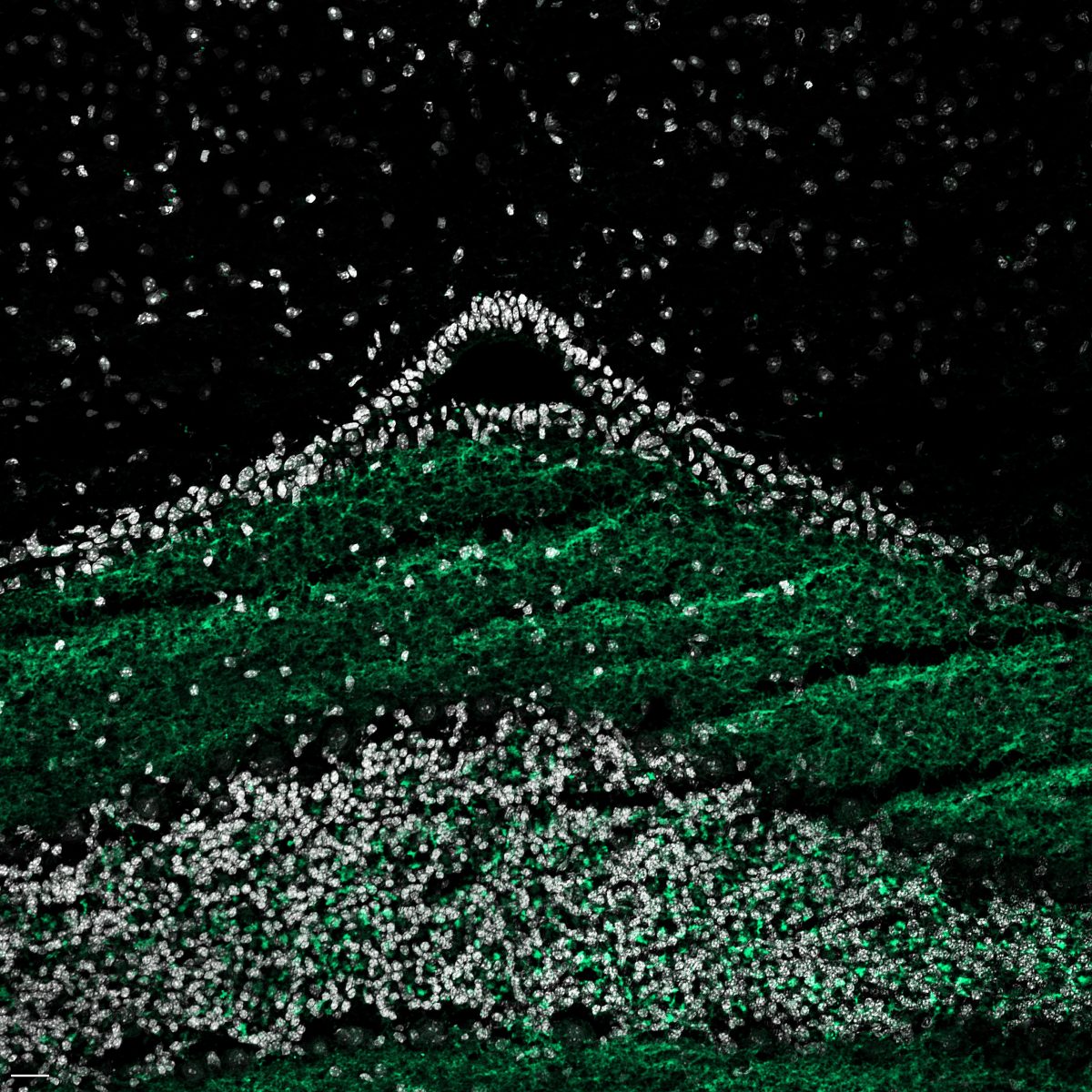
4. 37th View of Mount Fiji
Dr Kseniia Bondarenko, University of Edinburgh
You look at the coronal section of the mouse brainstem. The objects forming the top of the “mountain” are ependymal cells of the fourth ventricle’s choroid plexus, responsible for the production of cerebrospinal fluid (CSF). The fourth ventricle is one of the four connected CSF-filled cavities within the brain. Its brainstem part is seen here as a black space under the “snow cap.” The cerebellum occupies the bottom part of the image, below the layer of ependymal cells. Vesicular glutamate transporter 1 (anti-vGlut1 + AlexaFluor 488, green) is a sodium-dependent phosphate transporter responsible for uploading glutamate into synaptic vesicles. In the cerebellum, it localizes to the molecular layer. The granular layer is identifiable by the thick stratum of nuclei of the granular cells (Hoechst 33342, white). Prep: frozen 12 µm sections from CBA WT mouse, P19. The single optical section was acquired using Zeiss 980 confocal using 10x objective. Scale bar: 20 µm.
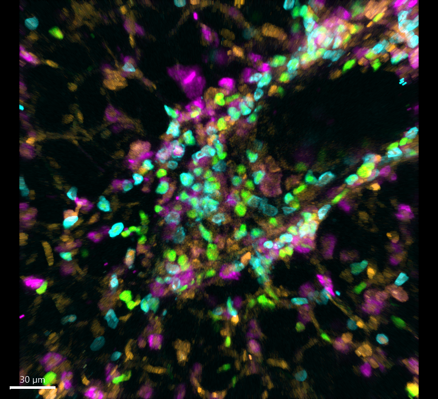
5. Multiplex Christmas Bauble
Dr Rachel Pennie, Scientific Officer CRUK Beatson Institute
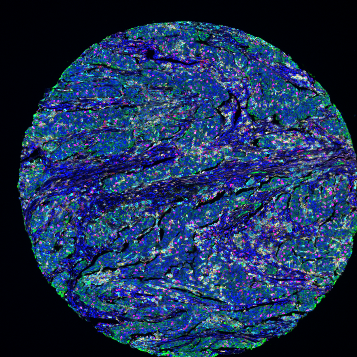
IF IHC multiplex panel on our lung adenocarcinoma cohort consisting of: CD4 (opal 570) – yellow CD68 (opal 690) – cyan CD8 (opal 520) – red CKPAN (Opal 620) – green Ki67 (opal 650) – magenta SMA (opal 540) – light pink The image was scanned on the Akoya Vectra Polaris at 20x. We are in the M42 group
6. Let it snow, let it snow, let it snow
Dr Cristina Martinez Gonzalez, University of Edinburgh
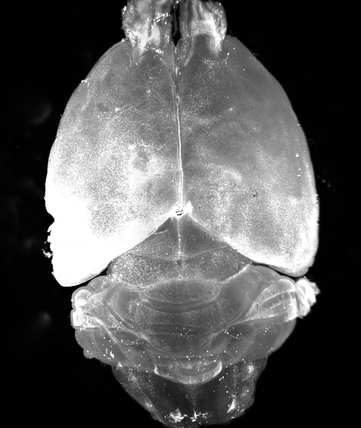
Light-sheet microscopy image of a whole mouse brain immunolabeled against cFOS (Alexa-647) and optically cleared using iDISCO. The animal used had a 50um saline injection in the left hippocampus. We imaged the brain with an Ultramicroscope II at 2X magnification in a single field of view. Optical slices were obtained every 4um across 3.5 mm in Z, covering the entire brain. A maximum intensity projection was the obtained, and adjusted for brightness and contrast in FIJI.
8. Cells Actin Like a Xmas Tree
Dr Jamie Whitelaw, University of the West of Scotland/CRUK Beatson Institute
NckAP1 KO MEFs stained with Vinculin (Red), Phalloidin (Green) and DAPI (Magenta). These were imaged on the Zeiss LSM 880 at the Beatson BAIR facility using the 63X oil objective lens. The images were processed to form an Xmas tree where the actin staining is the tree, the DAPI is baubles and the Vinculin as the Xmas lights.

Get in touch with the Scottish Microscopy Society!
Feel free to contact us or join our mailing list using the contact form below.


